Osteology Assessment: Report No 0304
Summary
In January 2004, York Osteoarchaeology Ltd was commissioned by Field Archaeology Specialists Ltd to carry out an osteological assessment of one human skeleton recovered during a watching brief at Nosterfield Quarry, North Yorkshire (NGR SE 7661 0886).
The skeleton had been interred in the partially backfilled ditch of a square barrow, provisionally dated to the Middle Iron Age. The barrow was part of a prehistoric landscape of ritual significance, which includes henge monuments, pit alignments, a cremation cemetery and further barrows.
Osteological analysis revealed that the skeleton was that of a man in his late thirties to mid forties. The bones of the arms, thighs and neck exhibited evidence for muscular strain indicative of their use in occupational activities. The man had also suffered from inflammation of the left leg, although this was healing at the time of his death. Two successive fractures of the left forearm would have caused disability of the left arm, which resulted in the subsequent greater use of the right arm in order to compensate. It is probable that these fractures were defence or work-related injuries.
 |
 |
 |
| Working shot, excavation of skeleton | Working shot, excavation of skeleton | Post excavation shot of skeleton |
Acknowledgements
York Osteoarchaeology Ltd would like to thank Field Archaeology Specialists Ltd for their help and support during this project.
Introduction
In January 2004 York Osteoarchaeology Ltd was commissioned by Field Archaeology Specialists Ltd to carry out an osteological assessment of one skeleton excavated in December 2003 during a watching brief at Nosterfield Quarry, North Yorkshire (NGR SE 7661 0886).
The skeleton (C1754) had been interred in a supine semi-flexed position in an oval pit (F335) cut into the partly backfilled ditch of a square barrow (F320). The individual had suffered slight damage to the left knee and right foot when the barrow ditch was re-cut at a later date. The skeleton was orientated NNE to SSW and the skull leant on both hands, which were positioned under the left side of the face, as if asleep. One horse tooth was recovered from between the legs of the skeleton and this may represent a grave good, or may have accidentally found its way into the grave, when the barrow ditch was re-cut. The burial has been tentatively dated to the mid Iron Age on the basis of its style.
Aims & Objectives
The skeletal assessment aimed to determine age, sex and stature, as well as to record any pathological conditions from which the individual may have suffered. Additionally, an attempt was made to identify the possible reasons for the unusual position of the arms of this individual.
Methodology
The skeleton was analysed in detail, assessing the preservation and completeness, as well as determining the age, sex and stature of the individual (Appendix 1). All pathological conditions were recorded and described.
Osteological Analysis
Osteological analysis is concerned with the determination of the demographic profile of the assemblage based on the assessment of sex, age and stature, as well as measurements and non-metric traits. This information is essential in order to determine the prevalence of disease types and age-related changes. It is also crucial for identifying gender dimorphism in occupation, lifestyle and diet, as well as roles of different age groups in society.
Preservation
Skeletal preservation depends upon a number of factors, including the age and sex of the individual as well as the size, shape and robusticity of the bone. Burial environment, post-depositional disturbance and treatment following excavation can also have a considerable impact on bone condition. Preservation of human remains is assessed subjectively, depending on the severity of bone surface erosion and post-mortem breaks, but disregarding completeness.
Preservation was assessed using a grading system of five categories: very poor, poor, moderate, good and excellent. Excellent preservation implied no bone erosion and very few or no post-depositional breaks, whereas very poor preservation indicated complete or almost complete loss of the bone surface due to erosion and severe fragmentation.
The skeleton was moderately well-preserved, due to extensive bone loss of the spongy bones of the joints and spine, and the cranium was fragmented, but complete. Additionally, many of the bones had suffered a moderate degree of fragmentation and erosion, some of which may have been caused when the skeleton was disturbed during re-cutting of the barrow ditch. Loss of the spongy bones meant that the skeleton was 70% complete.
Minimum Number of Individuals
A count of the 'minimum number of individuals’ (MNI) recovered from a cemetery is carried out as standard procedure during osteological assessments of inhumations in order to establish how many individuals were represented by the articulated and disarticulated human bones (without taking the archaeologically defined graves into account). The MNI is calculated by counting all long bone ends, as well as other larger skeletal elements, such as the hip joints and cranial elements. The largest number of these is then taken as the MNI. The MNI is likely to be lower than the actual number of skeletons which would have been interred on the site, but represents the minimum number of individuals which can be scientifically proven to be present.
As expected, the count of major bone elements provided a MNI of one individual.
Assessment of Age
The determination of age relies on the development and degeneration of bones and teeth. Different stages of development and degeneration have been mapped using data gathered from individuals of known age (Cox 2000). Methods used to determine age rely on the preservation of the dentition and hips. They are most precise when used to assess the developing skeleton, due to the fact that the growth of bones and teeth follows a relatively predictable course up to the age of twenty-five. However, the degeneration of the skeleton, which is assessed according to the severity of wear on the teeth, hips and ribs, depends not only on the age, but also on the sex, occupation, lifestyle and health of the individual analysed. The effect of wear on the teeth and bones tends to vary increasingly with advancing age; as a result, age cannot be reliably determined macroscopically beyond forty-six years.
Age was divided into a number of categories, including foetus (up to 40 weeks in utero), neonate (around the time of birth), infant (following birth to 1 year), juvenile (1-12 years), adolescent (13-17 years), young adult (18-25 years), young middle adult (26-35 years), old middle adult (36-45 years) and mature adult (46+ years). Age was determined using standard ageing techniques, specified by Buikstra and Ubelaker (1994) and Scheuer and Black (2000).
The poor preservation of the hips meant that the age determination was based on less accurate criteria. The dental wear, long bone fusion, grade of cranial suture closure and degeneration of a tiny surviving fragment of hip joint (auricular surface) suggested that this individual was an old middle adult, aged between 36 and 45 years.
Sex Determination
Sex determination is a vital part of the analysis of human remains, because of the likelihood that different sexes followed different lifestyles as a result of varying occupations, childbearing, or other activities, which may have affected their health. Sex assessment relies on the presence of the skull and pelvis, the morphology of which are sexually dimorphic, as described by Mays (2000).
Sex determination relied solely on the cranial morphology, which consistently suggested that this individual was male.
Metric Analysis
Stature can only be established if at least one complete and fully fused long bone is present. In this instance, none of the long bones were intact, which meant that stature could not be established. Leg measurements were obtained from the femora and tibiae and were used to calculate robusticity indices. From front to back, the right femoral shaft was broad and flat, while the left femoral shaft was more rounded. The platycnemic index (robusticity index) of the tibiae was calculated in order to establish the degree of tibial shaft flatness. The tibiae of skeleton C1754 were of average shape.
Craniometric measurements could not be taken because the skull was extremely fragmented. As a result, the skull shape could not be established. However, it was possible to establish through the survival of the nasal part of the face that this individual had a very prominent nose.
Non-metric Traits
Non-metric traits are additional sutures, facets, bony processes, canals and foramina, which occur in a minority of skeletons and are thought to suggest diversity and familial affiliation between skeletons (Saunders 1989). The origins of non-metric traits have been extensively discussed in the osteological literature and it is now thought that while most non-metric traits have genetic origins, some can be produced by factors such as mechanical stress (Kennedy 1989) or environment (Turkel 1978).
A total of thirty cranial and thirty post-cranial non-metric traits were selected from the osteological literature (Buikstra and Ubelaker 1994; Finnegan 1978; Berry and Berry 1967) and the skeleton was scanned for these traits. Two cranial traits were recorded, including a mastoid foramen extrasutural and a precondylar tubercle. Both traits are thought to have genetic origins. The skeleton was also found to have hypotrochanteric fossae on both femora, as well as a third trochanter on the left femur. These traits have been attributed to mechanical stress, in particular to the main bottom muscle, gluteus maximus, and may therefore be activity-related.
Pathological Analysis
Pathological conditions can manifest themselves on the skeleton during life, especially when these are chronic or the result of trauma to the bone. The bone elements to which muscles attach can also provide information on muscle trauma and excessive use of muscles.
Degenerative Joint Disease
Degenerative joint disease (DJD) is caused by a number of factors, including hereditary predisposition, increasing age, endocrine stress and mechanical strain. Occupational stress often affects the joint itself, whilst age may induce marginal changes. These marginal changes (osteophytes) are characterised by additional bone formation, whereas pitting affects the actual joint surface.
Evidence for DJD was observed in the spine of this man, in the form of mild pitting on the vertebral body surfaces of the cervical (neck) vertebrae. It is not uncommon for individuals in the 36 to 45 year age group to suffer from mild manifestations of joint disease.
Infection
Evidence for non-specific infection was very common before the introduction of antibiotics and is frequently observed in populations derived from archaeological contexts. Inflammatory lesions on human bones can be indicative of infectious diseases, such as leprosy and syphilis, or of non-specific infection, such as varicose veins, leg ulcers or trauma to the shins. However, skeletal lesions are only produced if the infection is chronic and long-standing (Roberts and Manchester 1995, 125).
Evidence for infection was observed in the form of superficial (periosteal) inflammatory lesions on the left tibia of Skeleton C1754. Any manifestations present on the right tibia would have been eroded. The nature of the lesions suggests that the inflammation was receding.
Trauma
Occasionally, it is possible to infer soft tissue trauma from the bones, in the form of muscular or ligamentous trauma. This is expressed trough the formation of bony processes (enthesopathies) at ligament attachments. Additionally, it is possible to observe cortical defects at the site of muscle attachments, which are the result of constant micro-trauma and are usually activity-related (Hawkey and Merbs 1995, 334). Cortical bone excavations were noted at a number of muscle attachments of Skeleton C1754, including brachialis at the ulnar tuberosities, which is a muscle that flexes the forearm. The skeleton also showed evidence for muscular strain to gluteus maximus, the main muscle of the bottom (discussed above). The muscle extends and laterally rotates the hip joint and extends the trunk. Evidence for an enthesopathy was noted at the back of the skull, where the trapezius muscle attaches to the occipital bone. The muscle also attaches to the shoulders and to the spine in the form of a large trapezoid, and is responsible for elevating, adducting and depressing the shoulders. This muscle is easily strained, which may have been the cause for the enthesopathy.
The most dramatic skeletal manifestation of pathology was observed at the left ulna. This bone had suffered a fracture at the lower third of the shaft. The fracture had moved from the supero-lateral part of the shaft obliquely to the infero-medial side. The fracture was well-healed, but had mal-united, causing overlapping of the two broken bone ends by 19.9mm. This would have caused considerable shortening of the bone.
The bone had been fractured a second time, from the same origin of the first fracture, but moving at a lesser angle medially (Plate 1). It is thus probable that the bone was fractured twice at the same site, but the overlapped part of the bone had been too strong to re-fracture, causing the fracture path to move superiorly to the initial fracture site. Notably, the second fracture is not united, and the two bone ends have formed a false joint with an irregular, pitted surface. It is assumed that the rotational stress placed on the forearm prevented union of the two fractured bone ends.
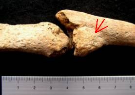
Plate 1. Left ulna showing overlapped healed fracture (arrow) and non-union of second fracture (centre)
Although the proximal and distal joints of the radius were missing, it cannot be ruled out that the bone was completely unaffected. Remarkably, the radius shaft was not fractured and the injury may have affected only the ulna.
Alternatively, it is possible that this fracture was a so-called 'Monteggia fracture’, which results in dislocation of the radial head (at the elbow joint) and fracture of the ulnar shaft, although the fracture site tends to be closer to the elbow.
It is certain that this injury would have hindered extensive use of the left forearm and it was therefore not surprising to note well-developed muscle attachments at the right humerus and right hand. It is possible that the initial injury was a parry injury, caused when the forearm is raised in front of the face or chest for protection against an attacker or advancing object. However, it is unusual for the bone to be broken twice at the same site, which may suggest that this was caused during a habitual activity. Alternatively, the second fracture may have been deliberately induced, with the aim of reducing the shortening of the ulna.
Dental Health
Analysis of the teeth of archaeological populations can provide vital clues about health, diet and oral hygiene, as well as information about environmental and congenital conditions.
Despite the poor survival of the upper jaw (maxilla), 27 of 32 teeth of this individual survived. The upper left second incisor and all four lower incisors were absent. The jaw bone holding these teeth was eroded and it was therefore not possible to determine whether the teeth had been lost ante-mortem or post-mortem. However, considering the lack of dental pathology, it is more likely that the teeth had been lost post-mortem. Loss of the anterior teeth in the burial environment is not uncommon, because the teeth are single-rooted and easily slip out of the jaw once the surrounding soft tissues have decayed.
Dental wear tends to be more common and severe in archaeological populations than in modern society, being caused by a much coarser diet based upon contemporary corn grinding techniques. Severity of the dental wear was assessed using a chart developed by Smith (1984). Each tooth was scored using a grading system ranging from 1 (no wear) to 8 (severe attrition of the whole tooth crown). The individual exhibited moderate to severe wear, which was consistent with his age.
Because of the lack of calculus deposits on the teeth, periodontal disease was slight and caused only mild resorption of the bone surrounding the teeth. The good dental health of this individual suggests that he was practising unusually good dental hygiene.
Mortuary Behaviour
The skeleton was interred in a ritually significant landscape, dating form the Neolithic to the late Iron Age. Nearby are the three Thornborough Henge Monuments, several pit alignments, a Bronze Age cremation cemetery, inhumations and round and square barrows. The burial was found in the ditch of a small square barrow. A second square barrow was located approximately 155m to the southwest of the burial, which may have been associated with a quadruple horse burial, 5m outside its perimeter.
The inhumation burials excavated at Nosterfield Quarry have so far proved most unusual. The two earlier burials represented secondary interments, following exposure of the body with subsequent loss of some bones and later burial of all surviving bones. While the burial of skeleton C1754 was not as remarkable, the position of his arms on the right side of the chest, with the hands positioned under the left cheek is very uncommon.
Discussion & Summary
The osteological analysis of a single skeleton from Nosterfield Quarry has provided a small glimpse into the life of this person. The skeleton was a male who had survived into his late thirties to mid forties. Poor skeletal preservation meant that it was not possible to establish his stature or obtain much evidence for facial characteristics, with the exception of the presence of a prominent nose.
This man enjoyed general good health, particularly of the teeth. He had, however, suffered from the effects of repetitive muscular injury to the arms and thighs, suggesting that he had carried out activities involving regular use of these muscles. Evidence for functional strain on the neck was noted in the form of muscular trauma, as well as mild degenerative joint disease on the vertebrae of the neck.
It is possible that trauma to the shins, an infectious disease or simply varicose veins were responsible for the development of inflammation on the left tibial shaft. The inflammation was receding, suggesting that it had begun to heal before the individual died. Inflammatory lesions to the shins are frequently observed in skeletons from all periods, but are usually more common in those populations from densely occupied urban areas.
Most notable, however, were two fractures of the left forearm, which may have been the result of a defence or work-related injury. The first fracture, which may have been associated with a dislocation of the radius at the elbow, resulted in considerable overlapping of the broken bone ends and must have caused considerable discomfort, as well as disabling the forearm. Whether the arm was fractured through another accident, or whether the second fracture was the result of an attempt to reduce the bone overlap could not be determined. Nevertheless, it is certain that the second fracture caused more discomfort as a result of the non-union of the broken bone ends. This resulted in greater use of the right arm, which was observed in the form of pronounced muscle attachments on the right upper arm and hand.
It could not be established, whether this injury was the reason for the unusual positioning of the arms of this man, which were placed on the left side of the chest, with his hands under the left side of his face, as if asleep. It is notable that this burial was disturbed during re-cutting of the barrow ditch, because this implies that those who worked on the ditch were not aware of the presence of the burial, or alternatively, that they were not afraid to disturb the burial of this man.
The presence of the horse tooth in the inhumation burial may suggest a tentative link with the quadruple horse burial near the larger barrow nearby. However, radiocarbon dating and further analysis of the archaeological evidence would be required to confirm any relationship between the burials.
Recommendations
It is recommended that the skeleton is subject to absolute dating, with the aim of establishing, whether this individual dates to the same period as the horse burial and inhumations previously excavated at Nosterfield Quarry. This may in turn aid the interpretation of the prehistoric landscape in which this man had been interred.
Once their date is known, focussed research on the mortuary rituals observed at Nosterfield Quarry could be considered, with the aim of establishing parallels for the funerary behaviour observed in the immediate locality of the site, and further afield.
References
Berry, A.C. and Berry, R.J. 1967. 'Epigenetic variation in the human cranium’, Journal of Anatomy 101 (2): 361-379
Buikstra, J.E. and Ubelaker D.H. (eds) 1994. Standards for Data Collection from Human Skeletal Remains (Fayetteville)
Cox, M. 2000. 'Ageing adults from the skeleton’, in M. Cox and S. Mays (eds), Human Osteology in Archaeology and Forensic Science (London): 61-82
Finnegan, M. 1978. 'Non-metric variation of the infracranial skeleton’, Journal of Anatomy 125: 23-37
Hawkey, D.E. and Merbs, C.F. 1995. 'Activity-induced musculoskeletal stress markers (MSM) and subsistence strategy changes among ancient Hudson Bay Eskimos’, International Journal of Osteoarchaeology 5: 324-338
Kennedy, K.A.R. 1989. 'Skeletal markers of occupational stress’ in M.Y. I_can and K.A.R. Kennedy (eds), Reconstruction of Life from the Skeleton (New York): 129-160
Mays, S. and Cox, M. 2000. 'Sex determination in skeletal remains’, in M. Cox and S. Mays (eds), Human Osteology in Archaeology and Forensic Science (London): 117-130
Roberts, C.A. and Manchester, K. 1995. The Archaeology of Disease (Stroud)
Saunders, S.R. 1989. 'Non-metric variation’, in M.Y. I_can and K.A.R. Kennedy (eds) Reconstruction of Life from the Skeleton (New York): 95-108
Scheuer, L. and Black, S. 2000. Developmental Juvenile Osteology (San Diego)
Smith, B.H. 1984. 'Patterns of molar wear in hunter-gatherers and agriculturalists’, American Journal of Physical Anthropology 63: 39-56
Turkel, S.J. 1989. 'Congenital abnormalities in archaeological populations’, in M.Y. I_can and K.A.R. Kennedy (eds) Reconstruction of Life from the Skeleton (New York): 109-127
Appendix A: Osteological & Palaeopathalogical Catalogue
| Skeleton Number | C1754 | |||||||||||||||
|---|---|---|---|---|---|---|---|---|---|---|---|---|---|---|---|---|
| Preservation | moderate | |||||||||||||||
| Completeness | 70%, the majority of the skull, long bones and hands are present | |||||||||||||||
| Age | 36-45, old middle adult | |||||||||||||||
| Sex | male | |||||||||||||||
| Stature | - | |||||||||||||||
| Non-Metric Traits | mastoid forarmen extrasutural (right), precondylar tubercle, hypotrochanteric fossae (bilateral), third trochanter (left) | |||||||||||||||
| Pathology | bone excavations for brachialis at ulnae, bone excavations for gluteus maximus at femora, enthesopathy for trapezius at occipital, double fracture of distal third of shaft of left ulna, well-healed with ma-union and overlap at first fracture, non-union at second fracture | |||||||||||||||
| Dental Health | 27/32 teeth present, 5 teeth lost pm, slight periodontal disease, moderate to severe wear | |||||||||||||||
| Right Dentition | Left Dentition | |||||||||||||||
| Present | p | p | p | p | p | p | p | p | p | - | p | p | p | p | p | p |
| Calculus | - | - | - | - | - | - | - | - | - | - | - | - | - | - | - | - |
| DEH | - | - | - | - | - | - | - | - | - | - | - | - | - | - | - | - |
| Caries | - | - | - | - | - | - | - | - | - | - | - | - | - | - | - | - |
| Wear | 4 | 6 | 8 | 7 | 6 | 6 | 7 | 7 | 7 | - | 6 | 6 | 7 | 8 | 6 | 4 |
| Maxilla | 8 | 7 | 6 | 5 | 4 | 3 | 2 | 1 | 1 | 2 | 3 | 4 | 5 | 6 | 7 | 8 |
| Mandible | 8 | 7 | 6 | 5 | 4 | 3 | 2 | 1 | 1 | 2 | 3 | 4 | e | 6 | 7 | 8 |
| Present | p | p | p | p | p | p | - | - | - | - | p | p | p | p | p | p |
| Calculus | - | - | - | - | - | - | - | - | - | - | - | - | - | - | - | - |
| DEH | - | - | - | - | - | - | - | - | - | - | - | - | - | - | - | - |
| Caries | - | - | - | - | - | - | - | - | - | - | - | - | - | - | - | - |
| Wear | 4 | 5 | 7 | 5 | 5 | 6 | - | - | - | - | 7 | 5 | 5 | 6 | 5 | 4 |
archaeological planning consultancy > thornborough > osteology assessment: report no 0304
Internet highlights
- UK Casinos Not On Gamstop
- Betting Sites Not On Gamstop
- Gambling Sites Not On Gamstop
- Non Gamstop Casino
- Best Non Gamstop Casinos
- Sites Not On Gamstop
- Casinos Not On Gamstop
- Best UK Casinos Not On Gamstop
- Non Gamstop Casinos
- Non Gamstop Casinos
- UK Sports Betting Sites
- UK Slot Sites
- Non Gamstop Casino
- Casinos Not On Gamstop
- Non Gamstop Casinos
- Non Gamstop Casinos
- Gambling Sites Not On Gamstop
- Casino Not On Gamstop
- UK Casinos Not On Gamstop
- UK Online Casinos Not On Gamstop
- Non Gamstop Casinos